40 diagram of the back muscles
25,583 back muscle anatomy stock photos, vectors, and illustrations are available royalty-free. See back muscle anatomy stock video clips. of 256. human musculature bodybuilding infographic muscular system vector human anatomy back muscle anatomy bicep male muscular anatomy human body anatomy female female anatomy muscle hamstrings muscle ... Deep muscles of the lower back include: The multifidus, a long muscle that travels nearly the entire length of the back.It helps to stabilize and rotate the lower back, and additionally takes some ...
The abdominal and lower back muscles work together to form a supportive "girdle" around your waist and lower back. Stronger muscles can help stabilize the lower back and can help reduce injury risk. 3. Stop smoking. Nicotine reduces blood flow to the spinal structures, including the lumbar discs, and can accelerate age-related degenerative ...

Diagram of the back muscles
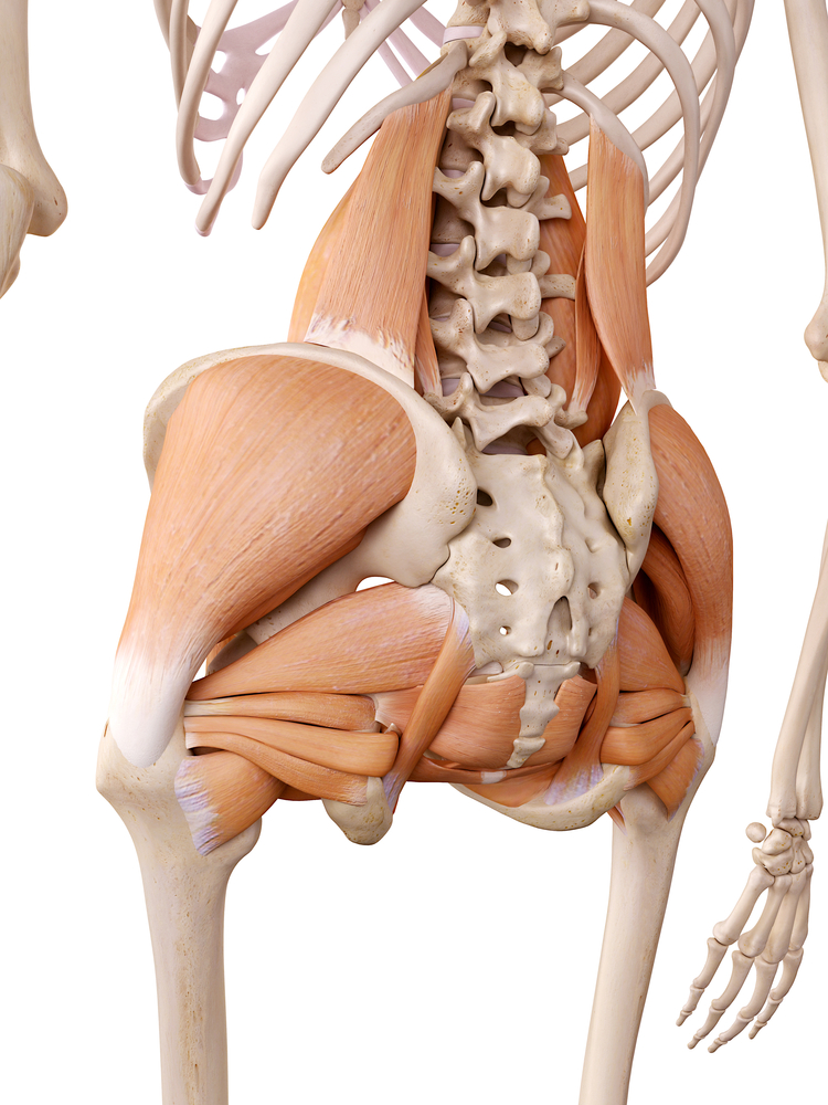
We are pleased to provide you with the picture named Anatomy Of Back Muscles Diagram.We hope this picture Anatomy Of Back Muscles Diagram can help you study and research. for more anatomy content please follow us and visit our website: www.anatomynote.com. Anatomynote.com found Anatomy Of Back Muscles Diagram from plenty of anatomical pictures on the internet. Muscle strain is often the cause of back pain from heavy lifting or vigorous exercise. But sometimes it's due to small jelly-filled disks meant to protect the space between vertebrae. When one ... The Quadratus Lumborum (QL) muscle, as seen in the diagram above, has a vital role in side bending your spine. The QL is a deep muscle that runs on both sides of the lower back. The muscle begins on the lowest rib (rib 12) and the nearby vertebra and connects to the pelvic crests.
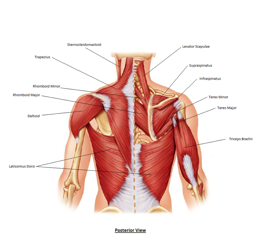
Diagram of the back muscles. Stretching exercises: Exercises to elongate and stretch your back muscles may include the knees to chest stretch or the prayer stretch.These stretches can improve the flexibility and mobility of your spine, providing for more freedom of motion. Strengthening exercises: Keeping your back muscles strong can help you recover from back injuries and may prevent future problems with your back. The signs and symptoms associated with active quadratus lumborum trigger points are as follows: Severe, deep, aching low back pain during movement or rest, and in nearly any position, but worse in the upright posture of standing or sitting. A sharp, knifelike pain when moving the hips/pelvis is common. Client’s will attempt to support and ... 740 lumbar spine anatomy diagram stock photos, vectors, and illustrations are available royalty-free. See lumbar spine anatomy diagram stock video clips. of 8. spinal vertebrae bone spine vertebra toracica spinal cord spine structure back diagram spine sections spinal cord vertebrae spinal structure health diagram. Try these curated collections. Muscular System Diagram Posterior (Back) View. by Sport Fitness Advisor Staff. This muscular system diagram shows the major muscle groups from the back or posterior view. To see a muscular system picture from the anterior (front) view click here. Occipitalis.
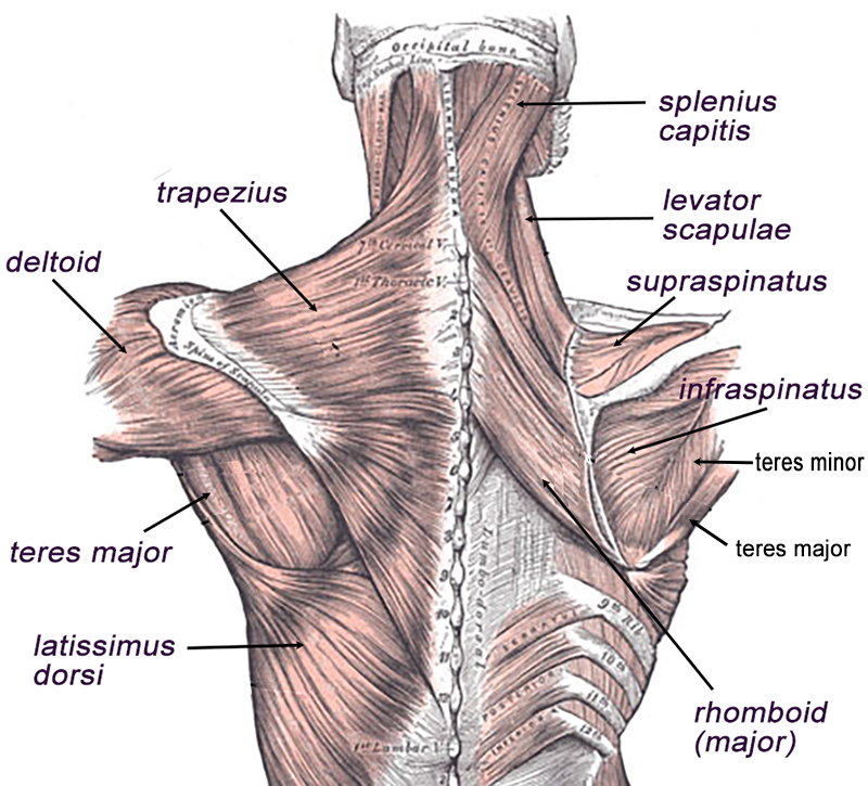
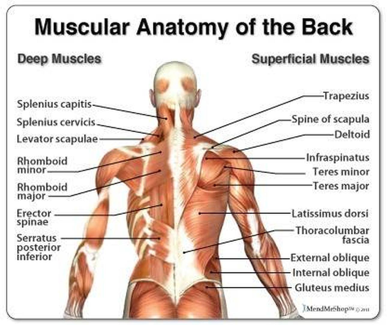
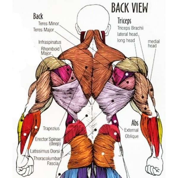
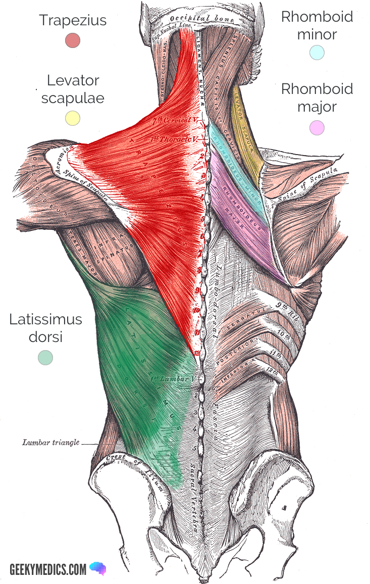
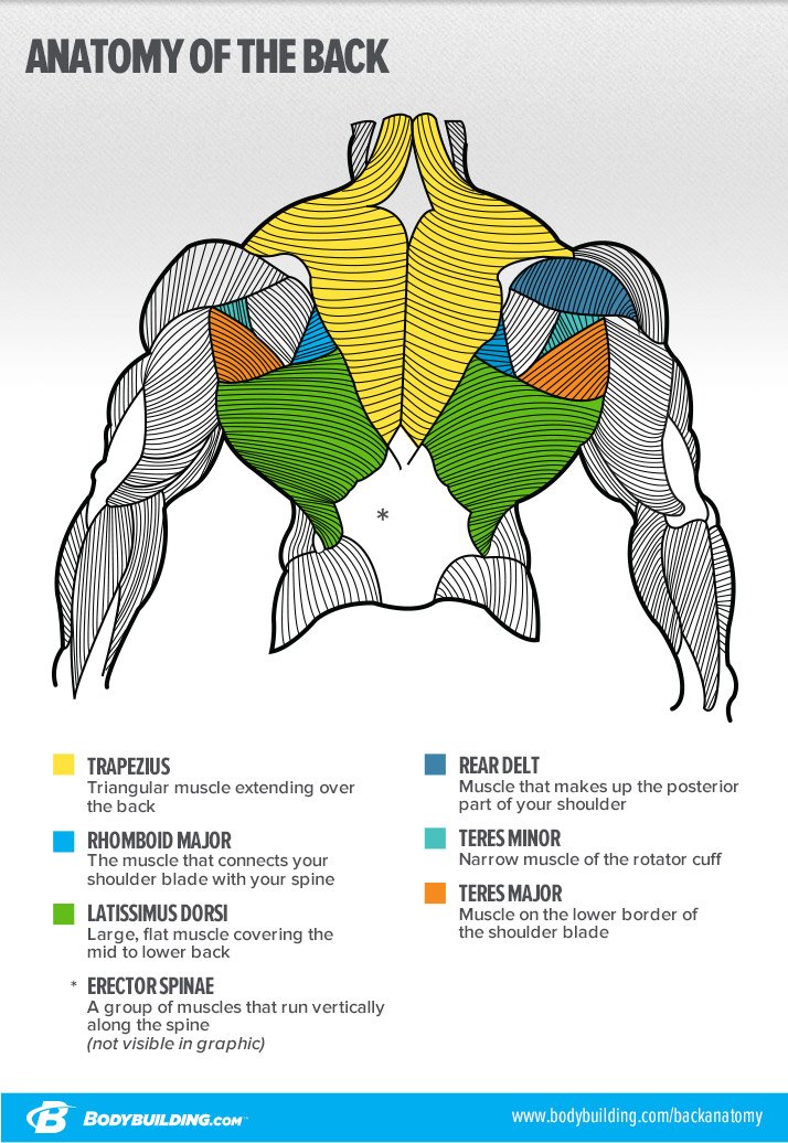
Back anatomy. The back is the body region between the neck and the gluteal regions. It comprises the vertebral column (spine) and two compartments of back muscles; extrinsic and intrinsic. The back functions are many, such as to house and protect the spinal cord, hold the body and head upright, and adjust the movements of the upper and lower limbs. Mar 05, 2018 · We have a collection of back muscle diagrams to help you learn more about the structure of muscles on your back. Three types of back muscles that help the spine function are extensors, flexors and obliques. The back anatomy includes the latissimus dorsi, trapezius, erector spinae, rhomboid, and the teres major. The following diagram below is the diagrams of back muscle. For your reference value these charts show the major superficial and deep muscles of the human body. For your reference value these charts show the major superficial and deep muscles of the human body. ... Claim your free copy of the client back care guide today. Your clients will thank you for it! Link to Client Back Care Guide. Links to FMA ... Jan 01, 2019 · Back Muscles: Names And Diagram. Daniel Nelson on January 1, 2019 2 Comments ! The human back extends from the buttocks to the posterior portion of the neck and shoulders. It is opposite from the chest, and the vertebral column runs down the back. The pelvis at the bottom of the back and the shoulders at the top of the back give the back its breadth, and it narrows in between these two regions.
Back muscles. The muscles of the back are a group of strong, paired muscles that lie on the posterior aspect of the trunk They provide movements of the spine, stability to the trunk, as well as the coordination between the movements of the limbs and The back muscles are divided into two large groups: The extrinsic (superficial) back muscles, which lie most superficially on the back. Anatomical diagrams of the spine and back. This human anatomy module is composed of diagrams, illustrations and 3D views of the back, cervical, thoracic and lumbar spinal areas as well as the various vertebrae. It contains the osteology, arthrology and myology of the spine and back. It is particularly interesting for physiotherapists ... Human muscle system, the muscles of the human body that work the skeletal system, that are under voluntary control, and that are concerned with movement, posture, and balance. Broadly considered, human muscle—like the muscles of all vertebrates—is often divided into striated muscle, smooth muscle, and cardiac muscle. Sep 04, 2019 · Muscles Of Lower Back Diagram. Muscles Of Lower Back Diagram. In this image, you will find an occipital bone, sternocleidomastoid, trapezius, deltoid in Muscles of the lower back diagram. As you can see, there are also have a spine of scapula deltoid, triceps brachii, latissimus dorsi. This picture also contains humerus, olecranon process of ulna, deep to tendon and so on.
Diagrams of Back Muscles | 101 Diagrams
Muscle layout and location shown in different colours/colors. 3d illustration. diagram of back muscles stock pictures, royalty-free photos & images. Human hand in front and back side. Hand in front and back side on isolated. Illustration about Human body part. diagram of back muscles stock illustrations.

Back Muscle Diagram / Muscles , 7 Deep Muscles Of Back ...
Back Muscles, Back Muscle Diagram. Creatine is now proving to be one of the most potent muscle growth accelerators giving excellent muscle mass increase and phenomenal strength increases order yours today. Creatine Research More Than a Sports Supplement Read More…. Some of the links in the post above are "affiliate links.".
Back Muscles Diagram Labeled : Female Muscle Diagram and ...
Trapezius. Postural and active movement muscle, used to tilt and turn the head and neck, shrug, steady the shoulders, and twist the arms. The muscle elevates, depresses, rotates, and retracts the scapula, or shoulder blade. Upgrade to remove ads. Only $2.99/month.

12 Back Exercises To Build A Stronger Better Back Help You ...
The longissimus (red, in the image above) are located between spinalis and the iliocostalis muscles. There are three sets of longissimus muscles: 1) above the cervical area (longissimus capitis), 2) in the cervical area (longissimus cervicis), and 3) in the upper back or thoracic area (longissimus thoracis) .

Human Back Bone Chart Back Bones Diagram Human Anatomy ...
The muscles of the lower back help stabilize, rotate, flex, and extend the spinal column, which is a bony tower of 24 vertebrae that gives the body structure and houses the spinal cord.The spinal ...

Djinn - 2018 Reference File
The muscles of the back can be arranged into 3 categories based on their location: superficial back muscles, intermediate back muscles and intrinsic back muscles.The intrinsic muscles are named as such because their embryological development begins in the back, oppose to the superficial and intermediate back muscles which develop elsewhere and are therefore classed as extrinsic muscles.
Back Muscle Anatomy Model - Human Anatomy
Three types of back muscles that help the spine function are extensors, flexors and obliques. The extensor muscles are attached to back of the spine and enable standing and lifting objects. These muscles include the large paired muscles in the lower back, called erector spinae, which help hold up the spine, and gluteal muscles.
Muscle and ligament pain in the lower back
Bonus Content: The 7-Day Back Pain Cure Back pain is one of the top reasons for missed work and second only to upper-respiratory infections for causing doctor visits. Most of the time, back muscle pain is diagnosed then "treated" with little more than a prescription of rest, painkillers and muscle relaxants.

Love Yoga Love Life: April 2013
Summary. The back consists of the spine, spinal cord, muscles, ligaments, and nerves. These structures work together to support the body, enable a range of movements, and send messages from the ...

Back Muscles Human Anatomy 1933
The back muscles enable you to stand up straight; support and protect your spine; and reach, pull and extend your arms and torso. Poorly developed back muscles lead to everything from muscle tweaks and pulls to imbalances of the musculature to the all-too-common hunched-over look (the "Neanderthal look").
Pictures Of Back Muscles
See Back Muscles and Low Back Pain. Nerves in your lower back. Five pairs of lumbar spinal nerves labeled L1 to L5 branch off your spinal cord and exit through small holes between the vertebrae. The part of the nerve that emerges out of the spine is called the nerve root.

tscblog: Training your back muscles for strength and power
Back Muscle Diagram With Lower Back Anatomy. This is a diagram of the larger and more surface muscles of the low back. The anatomy of the spine is complicated. To learn more, watch this VIDEO. Lower Back Muscle Diagram Anatomy Does Degenerative Disc Disease affect the Lower Back Muscles? Another common cause of lower back and hip pain is disc.

Flag
The Quadratus Lumborum (QL) muscle, as seen in the diagram above, has a vital role in side bending your spine. The QL is a deep muscle that runs on both sides of the lower back. The muscle begins on the lowest rib (rib 12) and the nearby vertebra and connects to the pelvic crests.

Ea: Anatomy of Diplostomia
Muscle strain is often the cause of back pain from heavy lifting or vigorous exercise. But sometimes it's due to small jelly-filled disks meant to protect the space between vertebrae. When one ...

An écorché with left arm extended to the side, seen from the back: diagram showing the outlines of the muscles. Line engraving by Campbell, 1816/1821.
We are pleased to provide you with the picture named Anatomy Of Back Muscles Diagram.We hope this picture Anatomy Of Back Muscles Diagram can help you study and research. for more anatomy content please follow us and visit our website: www.anatomynote.com. Anatomynote.com found Anatomy Of Back Muscles Diagram from plenty of anatomical pictures on the internet.
Anatomy of the back: Spine and back muscles | Kenhub

Anatomy Diagram 1 - Male Chest Muscles

Diagrams of Back Muscles | 101 Diagrams

Lower Back Muscles Diagram - Human Anatomy Diagram | Lower ...

Pin by Nishiki Heavens on Anatomy | Lower back muscles ...

Black Rabbit, Byker Farm, Ouseburn Valley, Newcastle Upon Tyne, Tyne & Wear, England.

Allow Me To Lend You A Hand Or Foot. Fascinating Human Muscular Anatomical Ephemera From 1920

Muscle Diagram Most Important Muscles Of An Athletic Male ...

Friends Will Be Friends, Cats, Chlorakas, Paphos, Republic Of Cyprus.

Muscles Of Lower Back Diagram
What You Can't See In The Mirror Can Hurt You - 7 Most ...

Labeled Anatomy Chart Of Male Back Muscles On White ...

1996 - Beach Tent

leftover-sonobe-dodekaugmented-dodecahedron.2

BUILDING ASTRAL BRIDGES...Spaceship Earth is a Holographic Being... It will be like gazing at a hologram in a painting. The picture looks a certain way in one moment, and it changes completely in the next.

The 6 Best Muscles to Self Massage for Instant Relief of ...

Lower Back Muscles Chart - Diagram Of Back Muscles Of The ...

Muscles Diagrams: Diagram of muscles and anatomy charts ...

44 best Muscles & Anatomy images on Pinterest | Physical ...
Lower Back Muscle Diagrams Labeled

Personal - Sniper Muscular Study
The Big Reasons Your Back Isn't Growing

L0034583 'Muscles Man', Pseudo-Galen, Anathomia; WMS 290

Tantra and Breathing

Fat Loss, Building Muscle & Staying Fit: Human Anatomy Diagram
0 Response to "40 diagram of the back muscles"
Post a Comment