37 ligaments in the thumb diagram
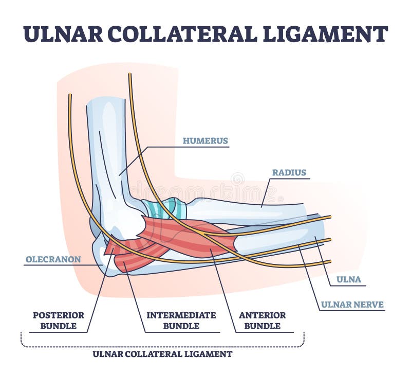
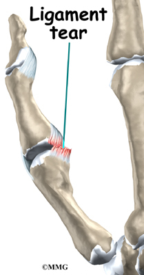
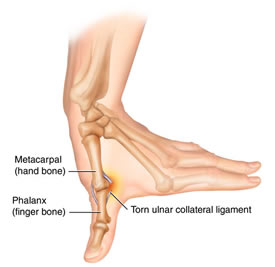
Interclavicular ligament. The sternoclavicular junction is the primary point of bony interaction between the upper axial skeleton and the upper limb. As such, the ligaments associated with this articulation will be discussed. The first to be encountered is the interclavicular ligament. A torn ligament in the thumb can be painful and an annoyance in everyday life. The technical term for a torn ligament in the thumb is an ulnar collateral ligament injury of the thumb. This type of injury occurs more often that most people would imagine. This type of injury is commonly referred to as the gamekeeper’s thumb or skier’s thumb. A torn ligament in the thumb can temporarily cause your hand to lose the ability to pinch and grasp objects. When there is a complete tear in the ligaments of the thumb, the metacarpophalangeal joint, or MCP joint is affected. This joint becomes weak and unstable, which causes the thumb to bend backwards.
Hand ligaments. The collateral ligaments of the hand are located on each side of all the fingers and thumb joints to limit the lateral movement of the fingers. The volar plate is the strongest ligament surrounding only the middle inter-phalangeal joints. It prevents the hyperextension of the fingers.

Ligaments in the thumb diagram
ligaments, previously as many as 16 ligaments identified (Bettinger) • Dorsal: Dorsal radial ligament (DRL) Dorsal central ligament (DCL) Posterior oblique ligament (POL) • Volar: Anterior oblique ligament (AOL) Ulnar collateral ligament (UCL) • Ulnar: First dorsal trapeziometacarpal ligament (DTM 1) Intermetacarpal ligament (IML) Collateral ligaments. The collateral ligaments course on either side of each interphalangeal joint, arising from the head of the more proximal phalanx and extending to the palmar, or volar, aspect of its distal counterpart. Arising from each collateral ligament is an accessory ligament, which extends anteriorly to attach to the fibers of the palmar ligament. The CMC joint of the thumb is located at the junction point of the thumb and the wrist. Break down the words in the name, carpometacarpal, and you get carpo- (wrist) and metacarpal (hand bone). This joint is commonly affected by arthritis. The CMC joint’s main function is to allow the thumb to open and grasp wide objects, like a basketball or ...
Ligaments in the thumb diagram. Collectively, the carpal bones form an arch in the coronal plane. A membranous band, the flexor retinaculum, spans between the medial and lateral edges of the arch, forming the carpal tunnel.. Proximally, the scaphoid and lunate articulate with the radius to form the wrist joint (also known as the 'radio-carpal joint'). In the distal row, all of the carpal bones articulate with the ... the thumb metacarpal52), and idio-pathic or hormonal-based laxity of the ligaments. Persons who require medical at-tention for instability of the thumb CMC joint often present with a combination of symptoms, which typically involves ligamentous laxity and resultant instability, pain, and functional limitations. These symp-toms may be precursors to An extension at the bottom of the radius where the wrist’s thumb-side collateral ligament attaches. A cashew-shaped bone close to the thumb that links the wrist’s two rows of carpal bones. A crescent-shaped bone located in the middle of the first of two rows of carpal bones comprising the wrist. A pyramid-shaped bone close to the pinky in ... Thumb Ligament Injuries: Skier's Thumb & Gamekeepers Thumb. A skier's thumb injury occurs when one falls against a planted ski pole, which hyperabducts the UCL. A gamekeeper's thumb occurs over time due to the motion Scottish fowl hunters use with their thumb and index fingers to break the head of a captured bird.
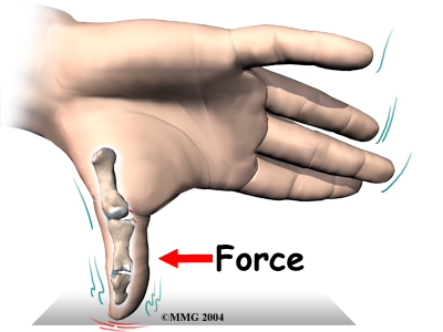
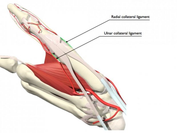
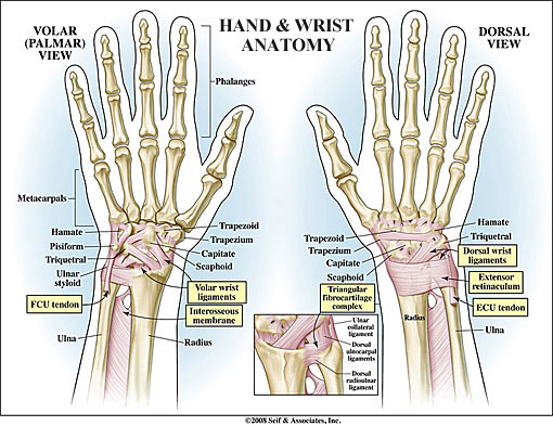
Thumb ligament injuries usually occur from a forced radial deviation (abduction) of the thumb during a high-velocity activity. Skiers and those who play ball-handling sports, such as baseball, football and basketball, have a greater risk of sustaining such an injury. Skiers can injure the thumb ligament in accidents involving the ski poles or ... When the ulnar collateral ligament of the thumb is injured, the MCP joint becomes painful and swollen, and the thumb feels weak when you pinch or grasp. You may see bruise-like discolorations on the skin around the joint. The loose end of the torn ligament may form a bump that can be felt along the edge of the thumb near the palm of the hand. Hand and Wrist Anatomy. The hand and wrist are made up of many different bones, muscles and ligaments that enable a wide range of movements. Bones. The following are the main structures of the hands: The wrist is formed where the two bones of the forearm - the radius (the larger bone on the thumb side of the arm) and the ulna (the smaller ... A torn ligament in the thumb usually affects the ulnar collateral ligament, which is used to pinch and grasp 3 4. The ulnar collateral ligament also stabilizes the thumb so it doesn't bend too far 3 4.An ulnar collateral ligament tear is often called a skier's thumb or a gamekeeper's thumb; skier's thumb describes an acute injury and gamekeeper's thumb a chronic injury, but the terms ...
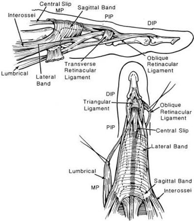
Jan 19, 2018 · The thumb joint has two collateral ligaments as well as the capsule, which is lined by a synovial membrane. The collateral ligaments are called the anterior and posterior ligaments. They are ... Anatomy of the Hand and Wrist: Bones, Muscles, Tendons, Nerves. The wrist links the hand to the forearm. The wrist is a complex system of many small bones (known as the carpal bones) and ligaments. The carpal bones are arranged in 2 interrelated rows. One row connects with the ends of the bones in the forearm—the radius and ulna. The thumb CMC joint has the most freedom of motion. The thumb metacarpal can bend and extend the thumb, move the thumb away from and toward the hand, and spin the thumb on the trapezium. Two very important ligaments are the dorsoradial and the volar beak ligaments. The abductor pollicis longus and brevis help move the thumb away from the hand. Ligaments are tough bands of tissue that connect bones together. Two important structures, called collateral ligaments, are found on either side of each finger and thumb joint.The function of the collateral ligaments is to prevent abnormal sideways bending of each joint. In the PIP joint (the middle joint between the main knuckle and the DIP joint), the strongest ligament is the volar plate.
My Thumb Winnipeg Cyclechick
Movements Of Thumb Diagram. Movements Of Thumb Diagram. Health care advices from Overseas Doctor . We are pleased to provide you with the picture named Movements Of Thumb Diagram. We hope this picture Movements Of Thumb Diagram can help you study and research. for more anatomy content please follow us and visit our website: www.anatomynote.com.

Basic Hand And Wrist Anatomy Hand Institute Of Charleston
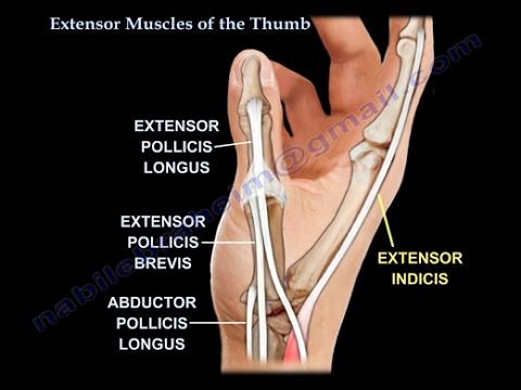
abduction of the thumb, a movement that takes place between the trapezium and the first metacarpal. • The extensor pollicis brevis. inserts at the base of the proximal phalanx of the thumb and is responsible for extension of the thumb. Additionally, because of its close relationship with the abductor longus, it helps in abduction.
Ligaments Of The Fingers Hand Orthobullets
Jan 21, 2018 · The thumb is the first of the hand's five digits, but it is typically not referred to as a finger. The thumb possesses a unique and wide range of motion not shared by the hand's other digits.

Skier S Thumb Physiopedia
The strongest ligament in the PIP joint is the volar plate. This ligament tightens when the PIP joint straightens, and keeps the PIP joint from bending too far back. Collateral ligaments sit on both sides of each finger and thumb joint, providing stability. These ligaments keep the finger joints from bending sideways.

Ligaments Of The Fingers Hand Orthobullets
Labelled Diagram of Phalanges of the Hand. ... Phalanges are bony basis of fingers and a place of insertions of ligaments and tendons. They create joints that together with corresponding muscles make finger flexion and extension possible. ... Hypoplastic thumb is a condition in which the osseous structures are hypoplastic, the thumb is ...
Ligament Reattachment With Suture Anchors For Proximal Phalanx Base And Shaft Skier S Thumb
Browse 321 hand anatomy tendons stock photos and images available, or start a new search to explore more stock photos and images. Vintage anatomical color illustration of the musculature of the human forearm. Hand, Illustration. Carpal Tunnel Syndrome, The Structures Of The Wrist Associated With Carpal Tunnel Syndrome.

Thumb Ligament Tear Skiier S Thumb Bone Talks
The radial collateral ligaments of the thumb MP are also injured at a higher rate than finger MP or IP joints but are less common than the ulnar collateral ligament. The radial collateral ligament is injured in forced adduction and leads to instability and eventually degeneration. If the ulnar collateral ligament is still intact, it also leads ...
Ulnar Collateral Ligament Sprain Skier S Thumb Stock Vector Illustration Of Injuries Medical 116064151
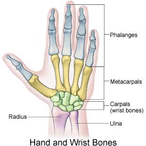
Anatomy of the Hand. The hand is composed of many different bones, muscles, and ligaments that allow for a large amount of movement and dexterity. There are 3 major types of bones in the hand itself, including: Phalanges. The 14 bones that are found in the fingers of each hand and also in the toes of each foot.

Injury To Ulnar Collateral Ligament Of Thumb Madan 2014 Orthopaedic Surgery Wiley Online Library
Thumb Joints The thumb consists of the first metacarpal and two phalanges. ... loosely proximally via two check rein ligaments, which fold allowing flexion (diagram 3). The main purpose of the volar plate is to provide stability by limiting ... Diagram 6: muscles of the thumb. 8 y and r e l e n n n n t s erus d s d e s e) d e a-f 5 th al d ar ...
Thumb Injuries Your Complete Guide To Diagnosing Thumb Pain
The ligaments of the female reproductive tract are a series of structures that support the internal female genitalia in the pelvis. The ligaments of the female reproductive tract can be divided into three categories: Broad ligament - a sheet of peritoneum, associated with both the uterus and ovaries. Uterine ligaments - ligaments primarily ...
Dorsoulnar Approach To The Mcp Joint Of The Thumb
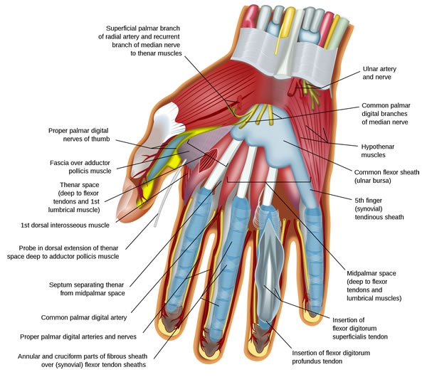
Part 1: Flexor tendon injuries Anatomy. There are two flexor tendons for each finger and one for the thumb. The flexor digitorum superficialis (FDS) and flexor digitorum profundus (FDP) are the flexor tendons of the fingers, and the flexor pollicis longus (FPL) is the only thumb flexor.. The flexor tendons travel distally from the forearm through the carpal tunnel and are named based on the ...
Physical Therapy In Miami Beach For Ulnar Collateral Lig Injuries Thumb
The CMC joint of the thumb is located at the junction point of the thumb and the wrist. Break down the words in the name, carpometacarpal, and you get carpo- (wrist) and metacarpal (hand bone). This joint is commonly affected by arthritis. The CMC joint’s main function is to allow the thumb to open and grasp wide objects, like a basketball or ...

Pediatric Trigger Thumb Ptt Symptoms Diagnosis And Treatment
Collateral ligaments. The collateral ligaments course on either side of each interphalangeal joint, arising from the head of the more proximal phalanx and extending to the palmar, or volar, aspect of its distal counterpart. Arising from each collateral ligament is an accessory ligament, which extends anteriorly to attach to the fibers of the palmar ligament.

Acute Finger Injuries Part I Tendons And Ligaments American Family Physician
ligaments, previously as many as 16 ligaments identified (Bettinger) • Dorsal: Dorsal radial ligament (DRL) Dorsal central ligament (DCL) Posterior oblique ligament (POL) • Volar: Anterior oblique ligament (AOL) Ulnar collateral ligament (UCL) • Ulnar: First dorsal trapeziometacarpal ligament (DTM 1) Intermetacarpal ligament (IML)
Sports Injuries To The Hand Wrist Elbow Ski Tennis Golf

Ulnar Collateral Ligament Of Thumb Wikipedia
Patient Education Concord Orthopaedics

Ligaments Of The Fingers Hand Orthobullets

Thumb Anatomy Mpc Joint Tendons Ligaments Hand Surgery Hand Health Hand Injuries
Thumb Anatomy Britannica

Sprained Thumb Hand Physiotherapy
Thumb Anatomy Ligaments Anatomy Drawing Diagram

Finger Tips Tendons And Ligaments Don T Forget The Bubbles
Wrist Anatomy New York Ny

Thumb Ulnar Collateral Ligament Injuries Florida Bone And Joint Specialistsflorida Bone And Joint Specialists
Thumb Ligament Injuries Physiopedia

Lateral Ulner Ligament Injuries Skier S Thumb Gamekeeper S Thumbs Stener S Lesion Knee Braces Ankle Supports Sports Injury Braces Thermoskin
Extensor Muscles Of The Thumb Everything You Need To Know Dr Nabil Ebraheim Youtube
Anatomy Of The Hand Giles Bantick Plastic And Hand Surgeon

Flexor Pulley System Hand Orthobullets

Disorders Ligament Injuries

Hand Ligament Anatomy Anatomy Drawing Diagram
Skiers Thumb London Hand Surgeon

Skier S Thumb Physiopedia
Body Anatomy Upper Extremity Tendons The Hand Society
Racgp Hands Fingers Thumbs Assessment And Management Of Common Hand Injuries In General Practice

Ulnar Collateral Ligament Sprain Ppt Download
0 Response to "37 ligaments in the thumb diagram"
Post a Comment