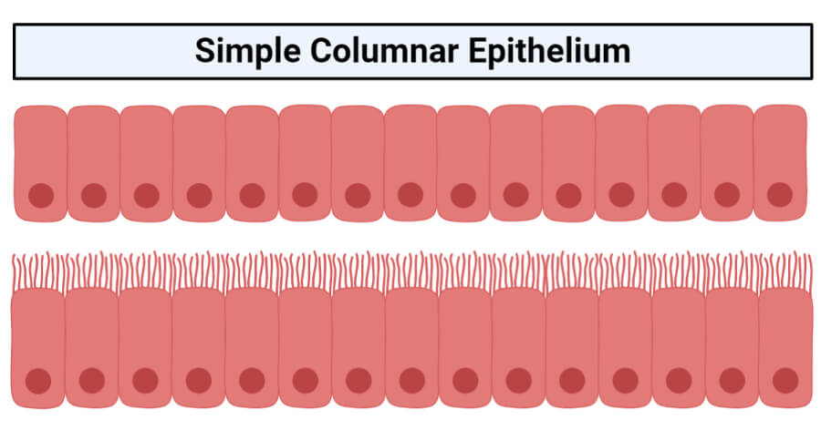
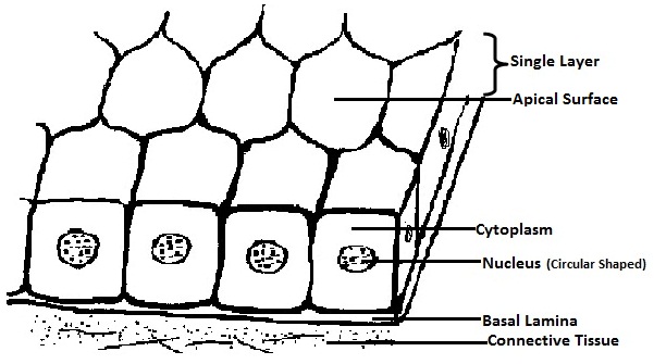
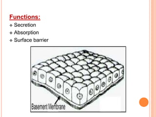
39 simple columnar epithelium diagram
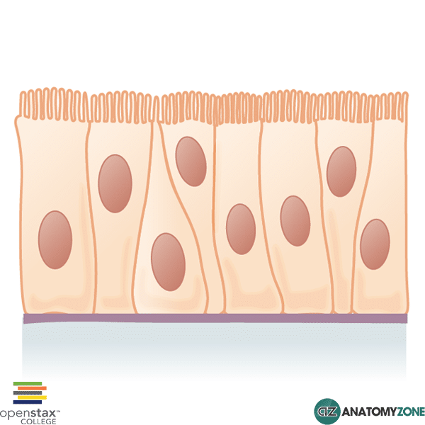
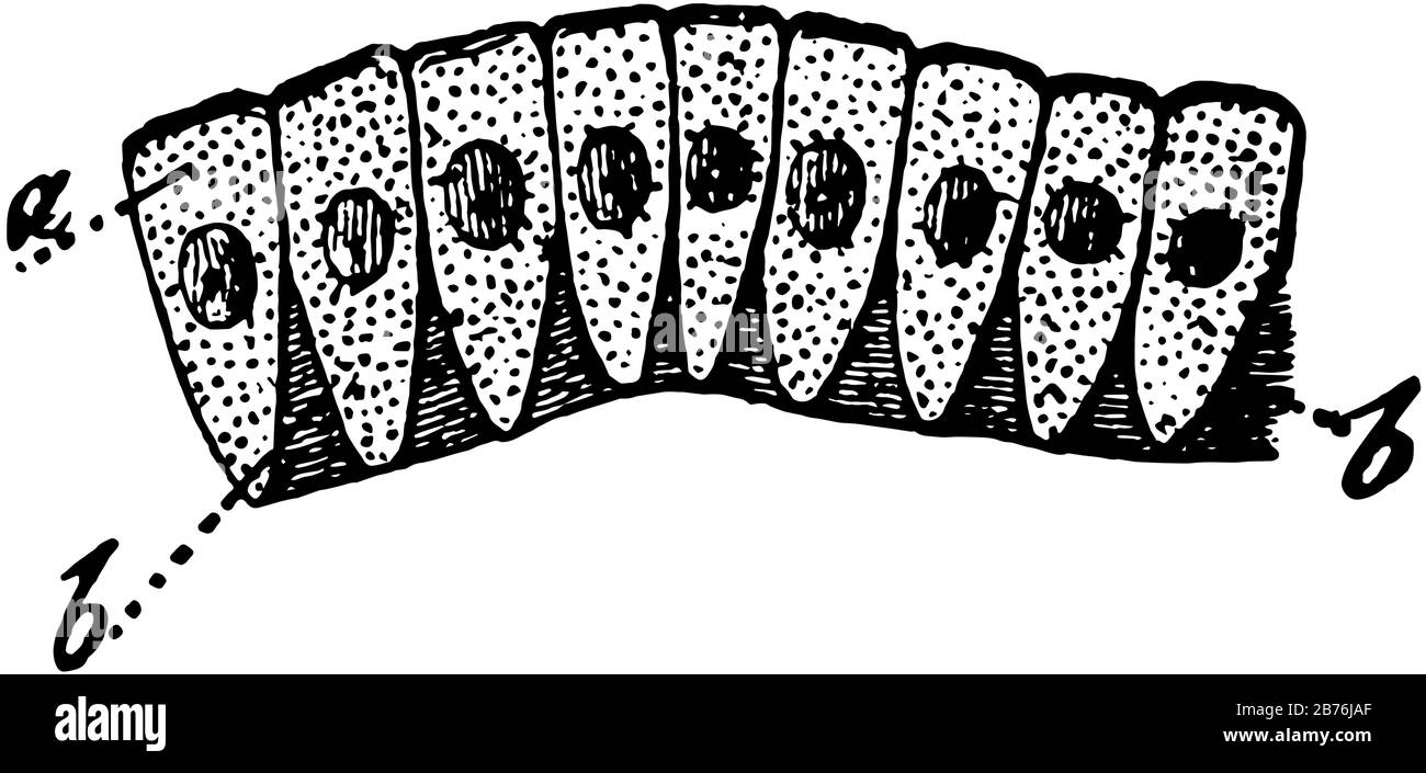
Simple columnar epithelium - Eugraph These absorptive cells are a single layer of columnar cells. (a simple columnar epithelium). Note an oval nucleus in the lower part of each columnar cell. Arrows indicate the base of this simple columnar epithelium sce. The lumen is indicated by lu. The surface area for absorption is increased by projections of the intestinal wall called villi. Simple Columnar Epithelium - Definition & Function ... Simple columnar epithelia are found in the stomach, small intestine, large intestine, rectum, fallopian tubes, endometrium, and respiratory bronchioles. In essence, they are found in parts of the respiratory, digestive and reproductive tracts where mechanical abrasion is low, but secretion and absorption are important.
Epithelial Tissue | histology A. Simple columnar epithelium. Slide 29 (small intestine) View Virtual Slide Slide 176 40x (colon, H&E) View Virtual Slide Remember that epithelia line or cover surfaces. In slide 29 and slide 176, this type of epithelium lines the luminal (mucosal) surface of the small and large intestines, respectively. Refer to the diagram at the end of this chapter for the tissue orientation and consult ...

Simple columnar epithelium diagram
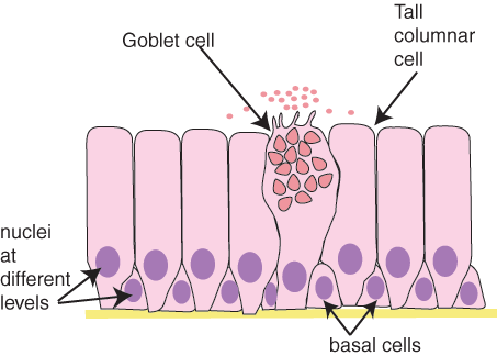
4.2 Epithelial Tissue - Anatomy & Physiology Simple columnar epithelium forms a majority of the digestive tract and some parts of the female reproductive tract. Ciliated columnar epithelium is composed of simple columnar epithelial cells with cilia on their apical surfaces. These epithelial cells are found in the lining of the fallopian tubes where the assist in the passage of the egg ... Simple Columnar Epithelium: A Labeled Diagram and ... Let's understand the structure of this type of epithelium with the help of the labeled simple columnar epithelium diagram given below. The nucleus of each of the cells is usually placed quite close to the thin, sheet-like basement membrane. Most of the nuclei are placed at the same level. Simple Columnar Epithelium Labeled Diagram Simple Columnar Epithelium Labeled Diagram Ciliated columnar epithelium is composed of simple columnar epithelial cells with cilia on their apical This illustration shows a diagram of a goblet cell. These labelled diagrams should closely follow the current Science (simple squamous epithelium). ORIGIN: columnar epithelium with goblet cells. TISSUE .
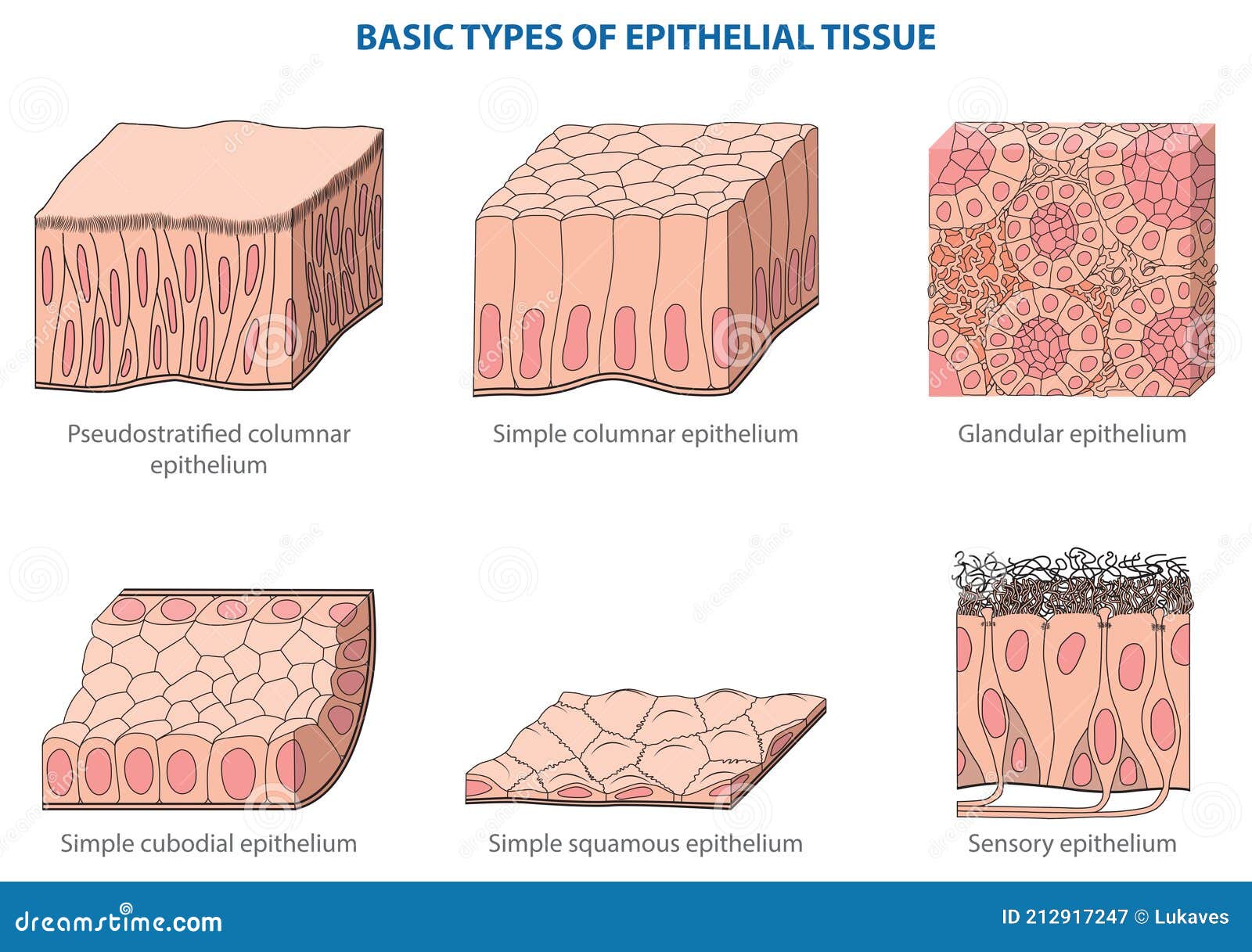
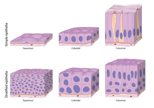
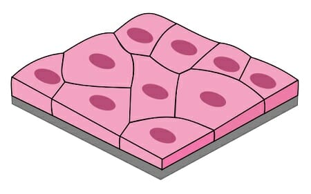
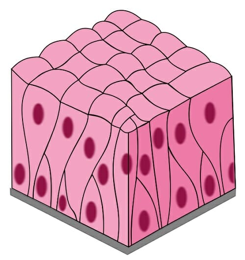
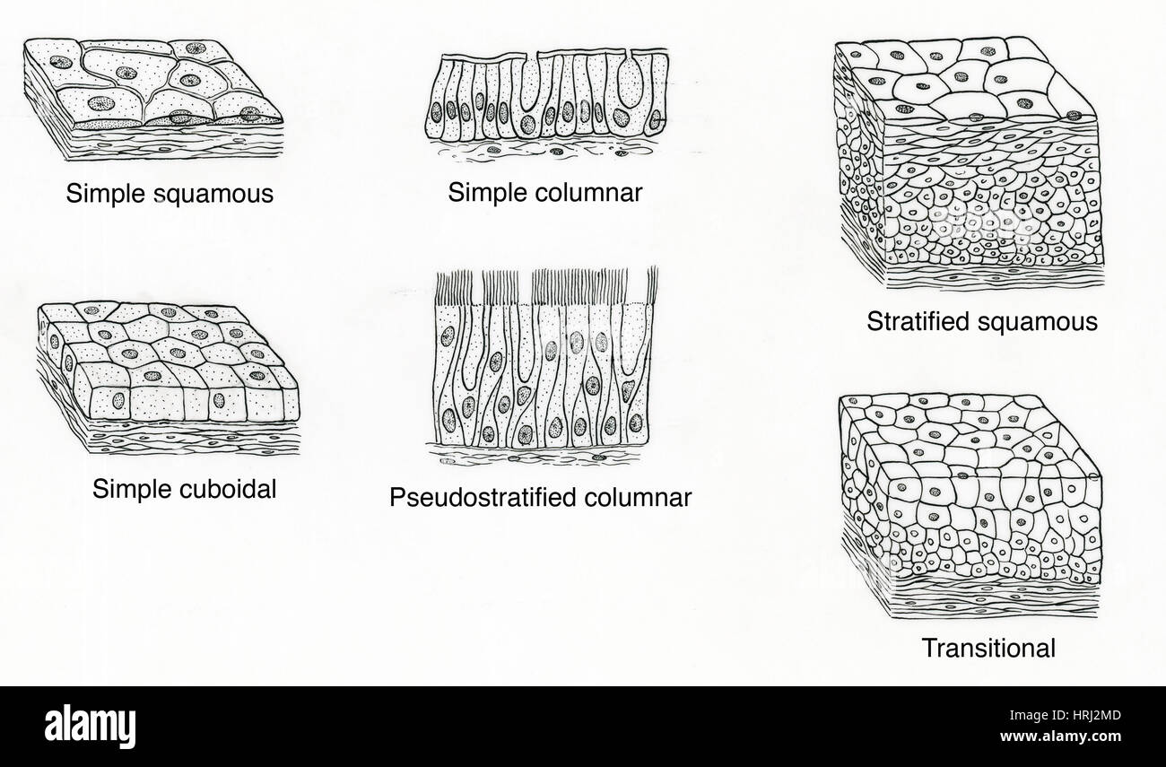
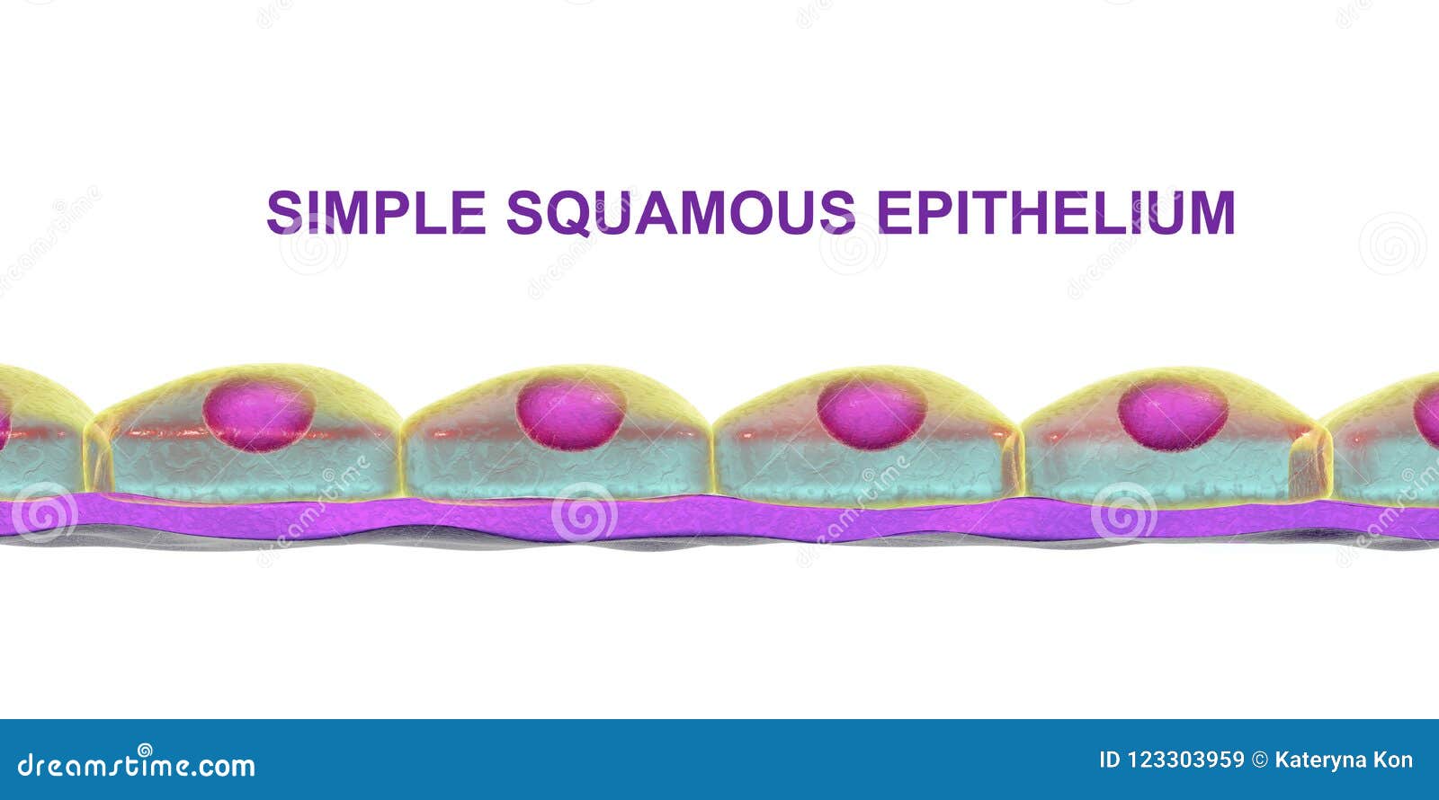
Simple columnar epithelium diagram. Simple Squamous Epithelium: Location and Diagram - Video ... The simple squamous epithelium location specifically exists in the lining of the blood vessels like the arteries, veins, and capillaries. It is also found lining the alveoli or air sacs within the... Simple columnar epithelium- structure, functions, examples The ciliated simple columnar epithelium is a single layer of ciliated column-like cells with oval nuclei near the base of cells, and the goblet cells are usually interspersed between the ciliated cells. The cilia are typically 5-10 μm long and 0.2 μm in diameter. Simple epithelium: Location, function, structure | Kenhub Synonyms: Simple columnar epithelium (with microvillous border) In this type of epithelium, the height of cells exceeds the width of the cell and seem closely packed narrow columns. The apical surfaces of these cells face the lumen and the opposite surface faces the basement membrane. The ovoid nuclei are usually placed towards basal surface. Epithelial Tissue-Introductory Diagrams and Images ... Ciliated Pseudostratified Columnar Epithelium (Diagram) Transitional Epithelium (Diagram) Simple Squamous Epithelial Tissue (Image) Stratified Squamous Epithelial Tissue (Image) Simple Cuboidal Epithelial Tissue (Image) Stratified Cuboidal Epithelial Tissue (Image) Simple Columnar Epithelial Tissue (Image) Stratified Columnar Epithelial Tissue ...
simple squamous epithelium - Epithelia: The Histology Guide This is a single layer of cells, and the cells are all tall columnar. diagram of columnar cells. Tall columnar epithelium lines the ducts of many exocrine ... Chapter 1, Page 10 - HistologyOLM - SteveGallik.org Diagram of a simple secretory columnar epithelium in section. A simple secretory columnar epithelium is found at two major sites in the animal body: 1) lining the stomach, and 2) lining the cervical canal. Our microscopic study of simple secretory columnar epithelium will consist of a study of the following specimen: Study #1. PDF Marieb HA8 chapter 4 - Pearson Simple Cuboidal Epithelium (Figure 4.3b) Simple cuboidal epithelium consists of a single layer of cube-shaped cells. This epithelium forms the secretory cells of many glands, the walls of the smallest ducts of glands, and the walls of many tubules in the kidney. Its functions are the same as those of simple columnar epithelium. Simple Columnar Epithelium (Ciliated) Diagram | Quizlet Start studying Simple Columnar Epithelium (Ciliated). Learn vocabulary, terms, and more with flashcards, games, and other study tools.
Epithelium Diagram | Quizlet Found underneath epithelia and above connective tissue. Made of basal and reticular lamina. It supports the overlying epithelia, provides a surface along which epithelial cells migrate across during growth and wound healing, and it acts as a physical barrier (to malignant melanoma) Basal lamina Secreted by the epithelial cells. Simple columnar epithelium - Wikipedia Simple columnar epithelium is a single layer of columnar epithelial cells which are tall and slender with oval-shaped nuclei located in the basal region, ... Epithelial Tissue: Structure with Diagram, Function, Types ... Columnar- long or column-like cylindrical cells, which have nucleus present at the base On the basis of the number of layers present, epithelial tissue is divided into the simple epithelium and stratified or compound epithelium Simple Epithelium- it is composed of one layer of a cell and mostly has a secretory or an absorptive function Simple Columnar Epithelium Labeled Diagram Simple Columnar Epithelium: A Labeled Diagram and Functions Epithelium is a tissue that lines the internal surface of the body, as well as the internal organs. Simple epithelium is one of the types of epithelium that is divided into simple columnar epithelium, simple squamous epithelium, and simple cuboidal epithelium.
PDF Week 1: Covering and Lining Epithelia These labelled diagrams should closely follow the current Science courses in histology, anatomy and ... EPITHELIUM: simple columnar or pseudostratified columnar TISSUE / ORGAN: seminal vesicle (ox) 1 2 blood vessel basal cell basement membrane ciliated columnar cell lamina propria
Simple columnar epithelium; stomach mucosa Diagram | Quizlet locations of simple columnar epithelium Nonciliated type lines most of the digestive tract (stomach to anal canal), gallbladder, and excretory ducts of some glands; ciliated variety lines small bronchi, uterine tubes, and some regions of the uterus. THIS SET IS OFTEN IN FOLDERS WITH... Simple Squamous Epithelium (serosa of th… 3 terms
Simple Columnar Epithelium Diagram - Quizlet Simple Columnar Epithelium Diagram | Quizlet Science Biology Histology Simple Columnar Epithelium STUDY Learn Flashcards Write Spell Test PLAY Match Gravity Created by sofievball_39 Terms in this set (14) How many cell layers are there in this tissue? One layer What is the shape of these cells? Tall columns / long-shaped
Simple Squamous Epithelium under a Microscope with a ... Simple squamous epithelium under a microscope consists of a single layer of thin, flat, and scale-like cells. These cells are joined together by an intercellular junction and rest on the basement membrane, whose thickness depends on the location. Here, I will show you what simple squamous looks like under a light microscope.
Ciliated Columnar Epithelium - Biology Self drawn diagram of ciliated columnar epithelium. Where is it? Ciliated columnar epithelial cells are found mainly in the tracheal and bronchial regions of ...
Simple Columnar Epithelium Labeled Diagram Simple Columnar Epithelium Labeled Diagram Ciliated columnar epithelium is composed of simple columnar epithelial cells with cilia on their apical This illustration shows a diagram of a goblet cell. These labelled diagrams should closely follow the current Science (simple squamous epithelium). ORIGIN: columnar epithelium with goblet cells. TISSUE .
Simple Columnar Epithelium: A Labeled Diagram and ... Let's understand the structure of this type of epithelium with the help of the labeled simple columnar epithelium diagram given below. The nucleus of each of the cells is usually placed quite close to the thin, sheet-like basement membrane. Most of the nuclei are placed at the same level.
4.2 Epithelial Tissue - Anatomy & Physiology Simple columnar epithelium forms a majority of the digestive tract and some parts of the female reproductive tract. Ciliated columnar epithelium is composed of simple columnar epithelial cells with cilia on their apical surfaces. These epithelial cells are found in the lining of the fallopian tubes where the assist in the passage of the egg ...



:watermark(/images/watermark_only_sm.png,0,0,0):watermark(/images/logo_url_sm.png,-10,-10,0):format(jpeg)/images/anatomy_term/simple-columnar-epithelium-4/PpLJVvrFTM3lZhGhVOhQTg_3Simple_columnar_epithelium.png)





:background_color(FFFFFF):format(jpeg)/images/article/en/simple-epithelium/VKbSAUnFnI6qqyq2cS0ag_qDDI8y5Bsv32llNotfSCA_Simple_columnar_epithelium_with_striated_border01.png)






0 Response to "39 simple columnar epithelium diagram"
Post a Comment