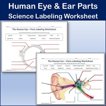
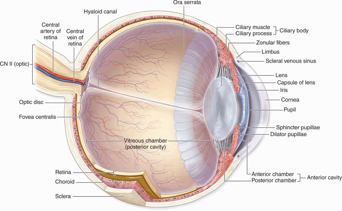
37 eye and ear diagram
At the bottom of the ear canal is the tympanic membrane which establishes the border between the external and middle ear. Auricle The auricle, also known as pinna, is a wrinkly musculocutaneous tissue that is attached to the skull and it functions to capture sound. The auricle is mostly made up of cartilage that is covered with skin.There are two aspects of the auricle: and medial (inner) and ...
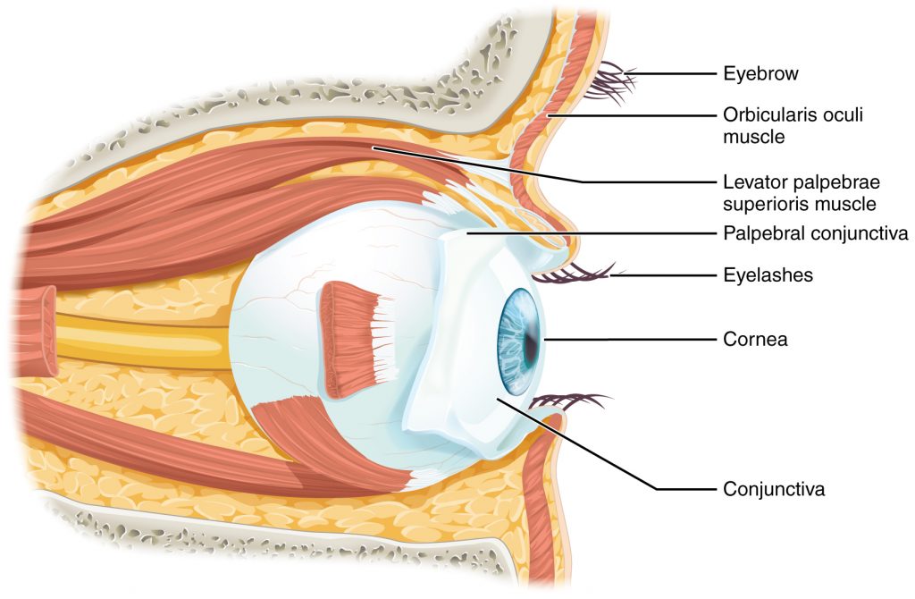
The eye is cushioned within the orbit by pads of fat. In addition to the eyeball itself, the orbit contains the muscles that move the eye, blood vessels, and nerves. The orbit also contains the lacrimal gland that is located underneath the outer portion of the upper eyelid. The lacrimal gland produces tears that help lubricate and moisten the ...
Ear model project. Learn about the amazing ears for kids by making this amazing, simple-to-make, ear model project.. This cleverear anatomy science project is perfect for preschoolers, kindergartners, grade 1, grade 2, grade 3, and grade 4 students to start to understand how the ear hears sounds.. Ear anatomy science project. We've been learning about the senses this week at our house.

Eye and ear diagram
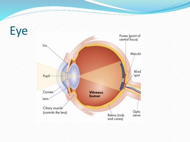
1) When we look at an object, then light rays coming from the object enter the pupil of the eye and fall on the eye-lens. 2) The eye-lens is a convex lens, so it converges the light rays and produces a real and inverted image of the object on the retina. 3) The retina has a large number of light sensitive cells.
The first turn of the canal bulges into the middle ear and is called the promontory; the outline of this can often be seen when viewing the tympanic membrane with a microscope. Peripheral vestibular system. The peripheral vestibular system is responsible for maintaining balance, coordinating the position of the head and eye movement.
The hoof is the foot of the horse, consisting of a hard exterior and softer interior. Similar to a fingernail but much stronger. Knee. The knee (carpus) is a large, bending joint of the front leg. It functions more like a wrist than a knee. Muzzle. The muzzle consists of the nose, mouth, and chin of a horse. Pastern.
Eye and ear diagram.
Embryology of the eye The eyes are formed from several embryonic layers. The epithelium of the cornea and lens are derived from the surface ectoderm.The endothelium of the cornea, sclera, and choroid arise from the neural crest cells.The neuroectoderm produces the posterior part of the iris, optic nerve, and retina. The remaining fibrous network and vasculature of the eye arise from the ...
Anatomy of the Eye · conjunctiva . · sclera , connecting the eyelids to the eyeball. · iris is the colored part of the eye. · pupil , the hole at the center of the ...Jan 26, 2021 · Uploaded by DrBruce Forciea
58,315 human eye anatomy stock photos, vectors, and illustrations are available royalty-free. See human eye anatomy stock video clips. of 584. eye vector outline anatomy of eye cartoon anatomy of the eye eye anatomy engraved shape anatomy of the eye eye nerve human engraving eye light waves diagram black white anatomy sketch.
View Adobe Scan Dec 3, 2021 (2).pdf from BSC 2093 at Indian River State College. 1. Parts of the ear: Match the part of the ear to its function and label its location on the diagram below. Eye and
Set of Human Sensory Organs: Anatomy of Five Sensory Organs, Eyes, Ear, Skin, Nose and Tongue. Sensory Organs provide us with data for perception, and it is the physiological capacity of all living organisms. Five sense organs are equipped in the human body. Those organs provide us with first-hand information about our external or internal world.
Eye Model Labeled Bing Images Eye Anatomy Anatomy Of The Eye Eye Health. Image Result For Eye Structure And Function Eye Anatomy Eye Anatomy Diagram Basic Anatomy And Physiology. Bio201 Eye Model Eye Anatomy Human Body Systems Projects Nervous System Anatomy. Image Result For Eye Model Labeled Eye Anatomy Anatomy Models Labeled Anatomy Models.
Take a moment to look at the ear model labeled above. This shows you all of the structures you've just learned about in the video, labeled on one diagram. Seeing them all together in this way is a great way to learn, since anatomical structures do not exist in isolation. That's why labeling the ear is an effective way to begin your revision.
The eye is the primary organ for sight, and the ear is the primary organ for sound ... Large Human Eye Anatomy Diagram Human Body Anatomy, Human Anatomy And ...
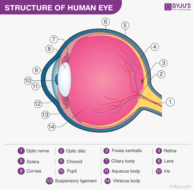
Eyeball (Bulbus oculi) The eye is a highly specialized sensory organ located within the bony orbit.The main function of the eye is to detect the visual stimuli (photoreception) and to convey the gathered information to the brain via the optic nerve (CN II).In the brain, the information from the eye is processed and ultimately translated into an image.
6,604 human eye diagram stock photos, vectors, and illustrations are available royalty-free. See human eye diagram stock video clips. of 67. diagram of the eye diagram of eyeball cross section of human eye section of human eye cross section eye cross section of eye anatomy of the eye eye parts human eye diagram of eye.
Start studying Psychology- Eye & Ear Diagram. Learn vocabulary, terms, and more with flashcards, games, and other study tools.
12,612 human ear anatomy stock photos, vectors, and illustrations are available royalty-free. See human ear anatomy stock video clips. of 127. the ear anatomy of ear ear anatomy the human ear anatomy of the ear ear diagram ear structure diagram of ear inner middle ear human ear parts. Try these curated collections.
Bony cavity within the skull that houses the eye and its associated structures (muscles of the eye, eyelid, periorbital fat, lacrimal apparatus) Bones of the orbit. Maxilla, zygomatic bone, frontal bone, ethmoid bone, lacrimal bone, sphenoid bone and palatine bone. Structure of the eye. Cornea, anterior chamber, lens, vitreous chamber and ...
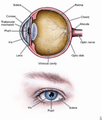
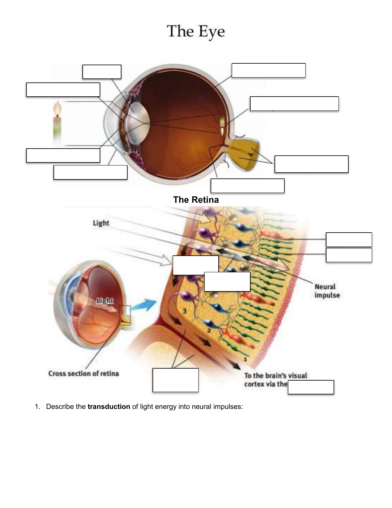
1. First, light passes through the cornea, a thin tissue that protects the eye and bends light to provide focus. 2. Next, light passes through the pupil, a small opening controlled by the iris. The iris is a colored muscle that constricts (gets smaller) or dilates (gets larger) based on light intensity. The more light exposure, the more the ...
They produce tears which help moisten the eye when it becomes dry, and flush out particles which irritate the eye. As tears flush out potentially dangerous irritants, it becomes easier to focus properly. Lens. The lens sits directly behind the pupil. This is a clear layer that focuses the light the pupil takes in.
Eye, Ears, and Sleep Disorders Nursing Test Banks. This section includes the NCLEX-style practice questions about eye, ears, nose, throat, and sleep disorders. This nursing test bank set includes 100 practice questions divided into two parts. ADVERTISEMENTS. Quizzes included in this guide are: Eyes, Ears, and Sleep Disorders Quiz #1 (50 Questions)
The eye area has some layers of skin that protects it. While the eyelids, eyelashes, and other parts of the eye area work together to make sure the eyes are moist, protected and functioning well, we should also make sure that the skin around the eyes is protected. This area is also quite sensitive and delicate, which means that it is vulnerable ...
The internal ear is a maze of bony chambers, called the bony, or osseous, labyrinth, located deep within the temporal bone behind the eye socket. Subdivisions. The three subdivisions of the bony labyrinth are the spiraling, pea-sized cochlea, the vestibule, and the semicircular canals.
Lesson 11. Handout 26. Diagrams of the Eye and Ear. Correctly label the diagrams according to the information your teacher gives you. Structure of the Eye.2 pages
The outer ear is the portion of the ear that sits atop the skull, which is made of flesh and cartilage. It is the visible part which serves to protect the eardrum. It also collects and guides sound waves into the middle ear. Compositional parts and their functions . Pinna (ear flap) The ear flap or pinna is the outer portion of the ear.
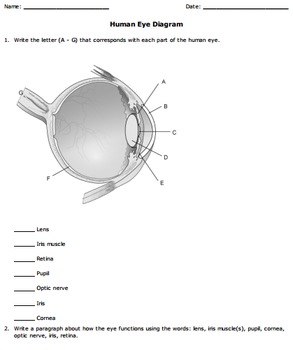
Human Eye Diagram: Contrary to popular belief, the eyes are not perfectly spherical; instead, it is made up of two separate segments fused together. Explore: Facts About The Eye To understand more in detail about our eye and how our eye functions, we need to look into the structure of the human eye.
The sensory cranial nerves are involved with the senses, search as sight, smell, hearing, and touch. Whereas the motor nerves are responsible for controlling the movements and functions of muscles and glands, cranial nerves supply sensory and motor information to areas of the head and neck. One nerve, the vagus nerve, extends beyond the neck to ...
The impulses are then processed by other neurons in the eye and by neurons in the brain. The vision center, the part of the cerebral cortex at the back of each ...25 pages
Labeled Eye Diagram Eye Anatomy Diagram, Human Eye Diagram, Diagram Of The ... Eye and Ear Models Eyeball Anatomy, Ear Anatomy, Sistema Visual, Eye Facts,.
In the diagram above - anatomy of the eye, the artery is shown in red while the vein is shown in blue. Tear Duct. This is a small tube that runs from the eye to the nasal cavity. Tear drains from the eyes in to the nose through the tear duct. This is why a teary eye is usually accompanied by a runny nose.
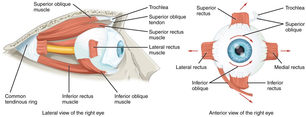
Eye Anatomy: Parts of the Eye Outside the Eyeball. The eye sits in a protective bony socket called the orbit. Six extraocular muscles in the orbit are attached to the eye. These muscles move the eye up and down, side to side, and rotate the eye. The extraocular muscles are attached to the white part of the eye called the sclera. This is a ...
The ear canal, or auditory canal, is a tube that runs from the outer ear to the eardrum. The ear has outer, middle, and inner portions. The ear canal and outer cartilage of the ear make up the ...
Students learn about the anatomical structure of the human eye and how humans see light, as well as some causes of color blindness. They conduct experiments as an example of research to gather information. During their investigations, they test other students' vision, gathering data and measurements about when objects appear blurry. These topics help students prepare to design solutions to an ...
Start studying Eye and Ear Disorders ch 43, 44. Learn vocabulary, terms, and more with flashcards, games, and other study tools.












0 Response to "37 eye and ear diagram"
Post a Comment